Artificial Intelligence in Medicine
Stroke Screening Support System
StrokeVista NCCT Acute (CMUH AIC)
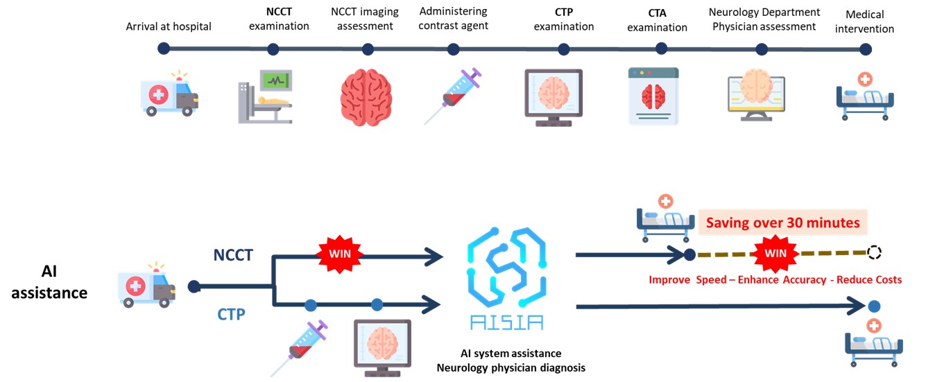
Ischemic stroke not only devastates patients' physiological functions but can also claim precious lives. Despite modern medicine's advanced computed tomography technology, the diagnostic process still faces severe challenges: standard non-contrast CT (NCCT) lacks sufficient sensitivity for acute ischemic stroke detection, while the more accurate CT perfusion (CTP) requires 15 to 30 minutes for scanning and contrast agent injection, potentially delaying critical treatment time. More problematically, not every hospital has CTP equipment and professional interpreters, and some patients cannot undergo this examination due to physiological factors, forcing physicians to make judgments based on limited information and inevitably increasing diagnostic uncertainty.
Our hospital's Artificial Intelligence Center, in collaboration with the Neurology Department, has successfully developed the "Non-contrast CT Ischemic Stroke AI Detection System," which can complete preliminary determination of acute ischemic stroke in just 90 seconds, while also visualizing brain-damaged areas in real-time and calculating ischemic volume! For acute large vessel occlusions with volumes greater than 70 milliliters, the system demonstrates excellent performance: 82% accuracy, 68% sensitivity, and 95% specificity, breaking through the limitations of traditional manual interpretation.
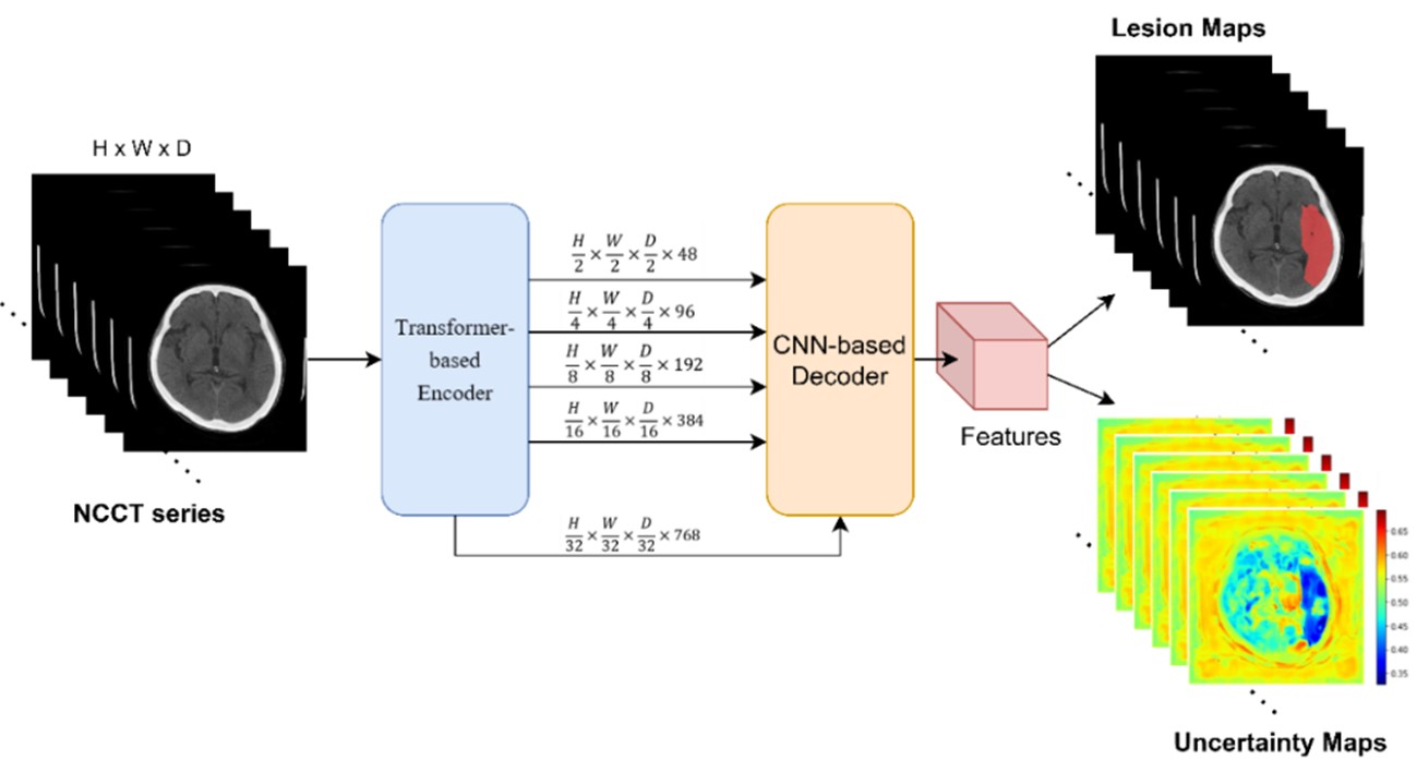
The system's greatest breakthrough is: it requires only a standard computed tomography (NCCT) without contrast agent injection to help physicians quickly identify large vessel occlusions (LVO) within 90 seconds! Not only does this save patients 30 precious minutes compared to traditional CTP examinations, but it also enables regional hospitals without CTP capabilities to make immediate interpretations and rapid transfer decisions, truly implementing zero time-lag in emergency and critical care!
Our hospital’s AI Center-developed “Non-contrast CT Stroke Detection System” has proven highly effective in identifying acute large vessel occlusions.
Journal Article
- Automated delineation of acute ischemic stroke lesions on non-contrast CT using 3D deep learning: A promising step towards efficient diagnosis and treatment.
https://www.sciencedirect.com/science/article/pii/S1746809424001976
Awards
- 2023 National Innovation Award – Academic Research Innovation Category
- Excellence Paper Award at the 23rd Annual Meeting of the Taiwan Neurological Society, and was also selected for presentation at the 2022 Asia Pacific Stroke Conference.
Ischemic Stroke Detection (EFAI)
According to global statistics, the annual incidence of intracerebral hemorrhage (ICH) is approximately 24 per 100,000 individuals. Notably, the incidence is higher among Asian populations, reaching 51.8 per 100,000 individuals.
This project employs deep learning algorithms to automatically analyze non-contrast head CT images of adults, identifying features indicative of acute ICH. Upon detection, the system issues alerts to the PACS/RIS workstations, assisting physicians in prioritizing cases for clinical evaluation and triage. The system accurately identifies the presence or absence of cerebral hemorrhage and classifies five types of intracranial bleeding: epidural hemorrhage, subdural hemorrhage, subarachnoid hemorrhage, intracerebral hemorrhage, and intraventricular hemorrhage.
Beyond rapid identification of suspected acute ICH, the system promptly notifies clinicians of critical findings, enabling timely intervention during crucial periods and facilitating earlier medical care for patients. The model demonstrates high diagnostic performance, with an accuracy of 98%, sensitivity of 95%, and specificity of 95%.
.jpg)
Patents
- TFDA medical device license (License No. 007937)
- U.S. FDA clearance (USFDA K231025).