Name of medical device
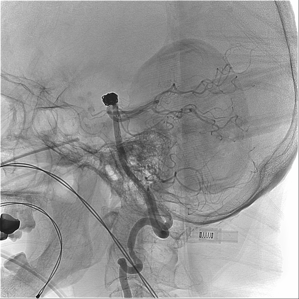
Vascular coil
Description
The coil is a metal medical device, which comes in various shapes, sizes and lengths. It can reduce blood flow and cause platelet agglutination by placing it in the diseased vessels, and then achieve embolization effects.
When to use
The coil can be used for embolization of bleeding vessels or aneurysms, which should be placed in the diseased vessels through the endovascular catheter.

Possible side effects
Contrast agent allergy, local wound bleeding, acute renal failure, acute pulmonary edema, arterial dissection, vasospasm, vascular rupture, stroke, and even death.
Postoperative care
- After the examination, the catheter is left at the blood vessel puncture site. Please avoid bending your thighs and take care not to pull it.
- If the catheter can be removed after evaluation, the doctor will apply pressure to the wound site to stop bleeding:
- Lie flat for 24 hours after hemostasis, and pressurize the puncture site with a sandbag for 8 hours.
- Family members are asked to pick up the sand bag every hour, observe whether there is bleeding or abnormality at the puncture site, and then press the sand bag back to its original place.
- If using styptic cotton or other hemostatic devices, sandbag pressure and flat lying time can be shortened. Please consult relevant medical staff.
- As a contrast agent is administered during the operation, it is advisable to drink more liquid and increase urination to expel the contrast agent from the body.
- During the first 2 weeks after discharge, avoid applying heavy pressure to the puncture site, do not carry heavy stuff or do strenuous exercise.
- Please receive follow-up exam regularly at the outpatient clinic after discharge.
- For an MRI exam, tell your physician or MRI technician that you have a vascular coil implanted for evaluation.

